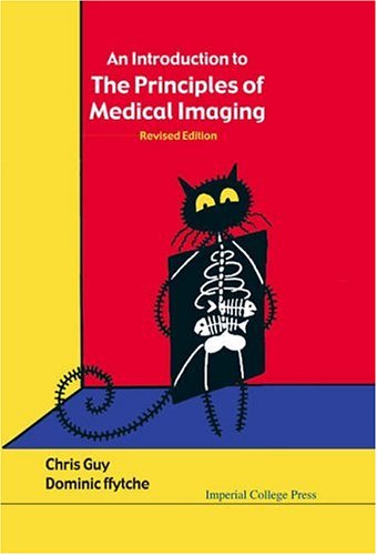
Chris Guy, Dominic Ffytche1860945023, 9781860945021, 9781860947469
This book is a non-mathematical introduction to the principles underlying modern medical imaging, taking tomography as its central theme. The first three chapters cover the general principles of tomography, a survey of the atomic and nuclear physics which underpins modern imaging, and a review of the key issues involved in radiation protection. The subsequent chapters deal in turn with X-ray radiography, gamma imaging, MRI and ultrasound. The clinical role of diagnostic imaging is illustrated in the final chapter through the use of fictional clinical histories. Three appendices provide a more mathematical background to the tomographic method, the principles of mathematical Fourier methods, and the mathematics of MRI.
This revised edition includes new introductory sections on the relevant physics of molecules in general, and water, in particular. Every chapter now has a table of key points with cross-references to other sections. Several figures have also been revised.
The book is intended to provide a broad introductory background to tomographic imaging for two groups of readers: the physics or engineering undergraduate thinking of specializing in medical physics, and the medical student or clinician using tomographic techniques in research and clinical practice.
Table of contents :
Contents……Page 28
Preface……Page 6
The Organisation of the Book……Page 9
Preface to the Revised Edition……Page 12
Acknowledgements……Page 14
SI Units……Page 16
Acceleration……Page 17
The First Law……Page 18
Angular Momentum……Page 19
Energy……Page 20
Field……Page 21
Planck’s Constant……Page 22
Pauli Exclusion Principle……Page 23
Contrast Enhancement……Page 24
Grey Scale……Page 25
Screening……Page 26
Voxel……Page 27
An Historical Background to Medical Imaging……Page 38
X-ray Radiography: The First Revolution……Page 40
X-ray CT: The Second Revolution……Page 41
MRI……Page 42
Diagnostic Ultrasound……Page 44
The Future……Page 45
1.1 Introduction……Page 48
Projections……Page 50
The Numbers in a Box Puzzle……Page 52
Simple Backprojection……Page 53
K-Space……Page 56
The Central Slice Theorem……Page 59
Digital Reconstruction……Page 60
Reconstruction Variations……Page 62
Digitisation of the Projections……Page 64
Signal to Noise Ratio……Page 65
Tomographic Image Artefacts……Page 66
Key Concepts in Chapter 1……Page 67
Questions and Problems……Page 68
2 Atomic and Nuclear Physics……Page 70
2.1 Introduction……Page 71
A Qualitative Survey of Medical Imaging Principles……Page 73
X-rays……Page 74
MRI……Page 75
Gamma Imaging……Page 76
The Need for Quantum Mechanics……Page 78
Photons — Quanta of Light……Page 79
Generating Light — Electromagnetic Waves……Page 80
The Electromagnetic Spectrum……Page 82
Atomic Structure……Page 83
The Bohr Atom……Page 85
Quantum Mechanics……Page 89
The Periodic Table of Elements……Page 90
Biological Atoms in the Periodic Table……Page 93
Molecules……Page 94
Some Properties of Water……Page 97
X-ray Scattering and Absorption by Atoms……Page 101
Elastic Scattering……Page 103
Inelastic Scattering……Page 104
Compton Scattering……Page 105
The Photoelectric Effect……Page 106
2.4 Nuclear Physics and Radioactivity……Page 108
Nuclear Structure……Page 109
Nuclear Binding Energy……Page 110
The Nuclear Shell Model……Page 111
Radioactive Decay……Page 113
Unstable Nuclei……Page 114
Radioactive Decay Law……Page 116
Natural Radioactivity……Page 117
Nuclear Fission……Page 119
Artificial Radionuclide Production……Page 120
Neutron Capture……Page 121
Particle Bombardment……Page 123
Atomic Magnetism……Page 124
Nuclear Magnetic Moments……Page 128
Nuclear Magnetic Resonance……Page 129
Key Concepts in Chapter 2……Page 134
Questions and Problems……Page 135
3.1 Introduction……Page 136
Exposure……Page 140
Absorbed Dose……Page 142
Dose Equivalent……Page 143
3.3 The Biological Effects of Radiation……Page 144
Stage 1 The Atomic Level……Page 146
Stage 2 Chemical Interactions……Page 147
Stage 3 Cellular and Whole Animal Changes……Page 148
Clinical Effects of Ionising Radiation……Page 149
3.4 Typical Medical Doses and Dose Limits……Page 150
3.5 Practical Radiation Protection and Monitoring……Page 152
External Protection Measures……Page 153
Dose Trends in Modern Diagnostic Techniques……Page 155
Ultrasound……Page 158
MRI……Page 159
Key Concepts in Chapter 3……Page 160
Questions and Problems……Page 161
4.1 Introduction……Page 162
4.2 The Linear X-ray Attenuation Coefficient……Page 165
Contrast……Page 169
Contrast Enhancement……Page 171
Scatter Reduction……Page 172
Detector Noise and Patient Dose……Page 173
Spatial Resolution and the Modulation Transfer Function……Page 176
X-ray Tubes……Page 178
X-ray Tube Operation and Rating……Page 181
The X-ray Spectrum……Page 182
Photon Detectors……Page 183
Photographic Film……Page 184
Film Characteristics……Page 185
Intensifier Screens……Page 188
Electronic Photon Detectors……Page 190
Ionisation Photon Detectors……Page 191
Scintillation Detectors……Page 195
The Image Intensifier……Page 197
4.5 The Modern X-ray CT Scanner……Page 199
The First Generation Scanner……Page 200
Third Generation Modern Scanners……Page 201
Hounsfield Units……Page 203
Partial Volume……Page 206
Patient Dose and Spatial Resolution in CT……Page 207
Standard Radiographs……Page 208
Angiography……Page 210
X-ray CT Images……Page 211
Key Concepts in Chapter 4……Page 213
Questions and Problems……Page 214
5.1 Introduction……Page 216
Carrier Molecules……Page 218
Radionuclides……Page 219
Technetium……Page 220
Iodine and Fluorine……Page 221
Xenon and Oxygen……Page 222
The Collimator……Page 223
The Scintillator Crystal……Page 224
XY Position and Energy Analysis……Page 225
The Inherent Low Count Rate in Gamma Imaging……Page 227
Gamma Imaging Source Response Functions……Page 229
5.5 Single Photon Emission Computed Tomography……Page 231
5.6 Positron Emission Tomography — PET……Page 234
The PET Detector……Page 236
True and False Coincidence Events in PET……Page 239
PET Correction Factors……Page 240
Images from Gamma Imaging……Page 242
Key Concepts in Gamma Imaging……Page 244
Questions and Problems……Page 245
6.1 Introduction……Page 246
Free Induction Decay……Page 249
T1 Relaxation……Page 252
T2 Relaxation……Page 254
A Microscopic Picture of Relaxation Mechanisms……Page 255
6.3 Pulse Sequences……Page 260
Saturation Recovery……Page 261
Spin Echo……Page 262
NMR Spectroscopy……Page 263
6.4 Spatially Localised NMR: MRI……Page 265
Slice Selection……Page 267
RF Pulse Shape……Page 268
Frequency Encoding……Page 272
Phase Encoding……Page 275
T1, T2 Weighting……Page 277
MR Contrast Agents……Page 280
6.5 Fast Imaging Methods……Page 281
Turbo Spin Echo……Page 282
Gradient Echoes……Page 284
Echo Planar Imaging……Page 285
Steady State Gradient Echo Methods……Page 287
3D or Volume Image Acquisition……Page 288
Time of Flight Methods……Page 289
Phase Contrast Methods……Page 291
6.7 Image Artefacts in MRI……Page 293
Static Field Distortions……Page 294
Time Varying Fields……Page 295
Water Fat Resonance Offset……Page 296
The Main Static Field……Page 297
Open Magnet Systems……Page 300
Gradient Coils……Page 301
The Radio Frequency Circuit……Page 302
Signal to Noise Ratio……Page 304
Images from MRI……Page 307
Image Artefacts……Page 310
Key Concepts in Chapter 6……Page 311
Questions and Problems……Page 312
7.1 Introduction……Page 314
The Velocity of Sound……Page 316
Ultrasound Radiation Field……Page 320
Attenuation……Page 321
Reflection of Ultrasound and Impedance — An Electrical Analogy……Page 322
Scattering……Page 323
Reflection……Page 324
The Ultrasound Doppler Effect……Page 326
Ultrasound Generation and Detection……Page 328
Standard B-Mode Imaging……Page 330
M-Scan or Time Position Plot……Page 331
Scanning Mechanisms……Page 332
The Electronic Amplification and Detection……Page 334
Spatial Resolution……Page 336
7.4 Doppler Imaging……Page 337
Miniature Invasive Ultrasound Probes……Page 339
Images from Ultrasound……Page 341
Key Concepts in Chapter 7……Page 342
Questions and Problems……Page 343
8.1 Introduction……Page 344
The Imaging Specialities……Page 345
The Medical Physicist and Radio-pharmacist……Page 346
X-rays……Page 347
Gamma Imaging……Page 349
PET……Page 351
MRI……Page 352
Ultrasound……Page 353
8.4 Clinical Examples……Page 354
Case 1 — X-ray, Fluoroscopy, CT, 99mTc -MDP Bone Scan……Page 355
Case 2 — CT, Fluoroscopy, MR, DSA……Page 356
Case 3 — MR, EEG, 18 FDG-PET……Page 357
8.5 Research Progress in Neuroimaging……Page 358
EEG and MEG……Page 362
PET Studies……Page 365
fMRI and the BOLD Effect……Page 367
Key Concepts in Chapter 8……Page 371
Questions and Problems……Page 372
A.1 Waves and Oscillations……Page 374
A.2 A Mathematical Description of Waves……Page 375
A.3 Fourier Analysis……Page 379
2D Fourier Analysis……Page 382
Sampling……Page 385
The Fast Fourier Transform……Page 387
Cleaning Up Noisy Images or Image Processing……Page 388
B.1 The Central Slice Theorem……Page 392
B.2 Filtered Backprojection……Page 394
Practical Filters……Page 397
C.1 Introduction……Page 400
C.2 Nuclear Moments in a Static Magnetic Field……Page 401
C.3 The Classical Picture of NMR……Page 403
Rotating Frames of Reference in Dynamics……Page 407
Bibliography……Page 412
Index……Page 416

Reviews
There are no reviews yet.