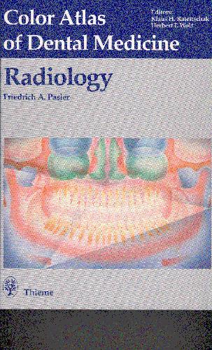
Pasler F.A., Hassell T.
Table of contents :
Cover Page……Page 1
Localization Using Various Methods……Page 2
Temporomandibular Joint Disturbances……Page 3
Index……Page 4
Radiographic Examination of the Patient in the Dental Office……Page 5
Examination Strategy and Active Protection Against Radiation Exposure……Page 6
Panoramic Radiography for Basic Information and Supplemental Examination Using Special Radiographs……Page 10
Technique for Panoramic Radiography……Page 12
Radiographic Anatomy in the Panoramic Radiograph……Page 28
The Bite-Wing Radiograph……Page 50
Apical and Periodontal Radiographic Technique……Page 54
Radiographic Technique with the Occlusal Film……Page 66
Radiographic Anatomy in Periapical and Occlusal Radiographs……Page 72
Localization Using Various Methods……Page 86
Errors in Technique That Reduce Radiograph Quality……Page 94
Processing Technique and Errors Leading to Poor Quality Radiographs……Page 106
Supplemental Examinations Using Conventional and Modern Imaging Techniques……Page 112
Selected Examples of Dental Radiographic Diagnosis……Page 128
Anomalies of Dental Development and the Teeth……Page 130
Concrements, Calcifications, Ossifications……Page 142
Regressive Changes in Teeth and Jaws……Page 150
Dentogenic Sinus Disorders……Page 162
Temporomandibular Joint Disturbances……Page 174
Cysts and Pseudocysts……Page 184
Odontogenic Tumors and Pseudotumors……Page 200
Nonodontogenic Tumors and Pseudotumors……Page 216
Traumatology……Page 250
Foreign Bodies and Postoperative Conditions……Page 258
Index……Page 268



Reviews
There are no reviews yet.