Table of contents :
Cover……Page 1
Preface……Page 2
How to approach cardiac diagnosis from the chest radiograph……Page 4
Frontal chest radiograph: normal border-forming structures……Page 5
Lateral chest radiograph: normal border-forming structures……Page 6
Skeletal abnormalities……Page 7
Questions to ask……Page 8
Left atrium……Page 10
Cardiac valve disease……Page 11
References……Page 12
Cardiac MR imaging techniques: general principles……Page 13
Cardiac MR imaging pulse sequences……Page 14
Scout images: transverse or axial plane……Page 15
Coronal and sagittal planes……Page 16
Short-axis plane……Page 17
Long-axis view through aortic and mitral valves……Page 18
Routine clinical studies with cardiac MR imaging……Page 19
Transposition of great arteries……Page 20
Tetralogy of Fallot……Page 21
Anomalous pulmonary veins……Page 22
Constrictive pericarditis versus restrictive cardiomyopathy……Page 24
Cardiac tumors and metastatic disease……Page 26
The expanding role of cardiac MR imaging……Page 27
Left ventricular function……Page 28
References……Page 29
Normal cardiac structures as seen on the plain film……Page 31
Enlargement of pulmonary artery segments……Page 33
Dilatation of the left atrial appendage segment……Page 35
Common causes of cardiac abnormalities seen on plain film in older adults……Page 36
Valvular heart disease……Page 37
Pericardial disease……Page 38
Adult congenital heart disease……Page 40
Plain film abnormalities in patients with lung cancer that suggest cardiac, pericardial, or large vessel involvement……Page 41
Postsurgical and posttraumatic abnormalities seen on plain films……Page 45
Other uses of MR imaging and CT in the thorax……Page 49
References……Page 51
Monitoring and therapeutic devices……Page 58
Venous……Page 59
Cardiac……Page 60
Atelectasis……Page 64
The cardiomediastinal silhouette……Page 65
Sternal dehiscence, osteomyelitis, and mediastinitis……Page 66
Pericardial complications……Page 67
Aortic pseudoaneurysm and dissection……Page 68
Coronary artery bypass graft surgery……Page 69
Aortic reconstruction……Page 70
Cardiac valve reconstruction and replacement……Page 71
Cardiac transplantation……Page 73
Congenital heart disease……Page 75
References……Page 77
Development of CT angiography……Page 80
Contrast-enhancement methods……Page 81
Postprocessing methods……Page 82
Phase-contrast imaging……Page 83
Dynamic contrast-enhanced MR angiography……Page 85
Atherosclerotic disease of the aorta……Page 86
Aortic aneurysm……Page 87
Aortic dissection……Page 88
Intramural hematoma……Page 91
Penetrating atherosclerotic ulcer……Page 92
Infectious and inflammatory aortic disease……Page 93
Adult congenital aortic disease……Page 95
Postsurgical evaluation of aortic diseases……Page 96
Traumatic aortic injuries……Page 97
References……Page 98
Functional anatomy……Page 101
Topographic anatomy……Page 102
Visualization of the pericardium……Page 103
Pericarditis……Page 104
Pericardial effusion……Page 105
Mechanical sequels of cardiac compression……Page 106
CT and MR imaging diagnostic criteria of pericardial constriction……Page 107
Morphologic types of pericardial constriction……Page 108
CT and MR imaging parameters of myocardial atrophy or fibrosis in pericardial constriction……Page 109
Postpericardiectomy hemodynamic and clinical results……Page 110
Summary……Page 112
References……Page 113
Calcifications of the heart……Page 116
Coronary artery calcification……Page 117
Myocardial calcification……Page 119
Pericardial calcification……Page 122
Valvular calcification……Page 124
Tumor calcification……Page 126
References……Page 128
Exercise or pharmacologic stress?……Page 131
One- or two-day protocol?……Page 132
Reporting of results……Page 133
Does gated single-photon emission CT add value?……Page 136
Does attenuation correction add value?……Page 139
Thallium 201 and technetium 99m sestamibi in assessing myocardial viability……Page 140
Nuclear perfusion imaging and the selection of patients for angiography……Page 142
References……Page 143
MDCT image acquisition……Page 147
Three-dimensional visualization……Page 149
Calcium scoring: clinical rationale……Page 151
Calcium scoring: technique……Page 153
Cardiac function……Page 154
MDCT coronary angiography: clinical rationale……Page 155
MDCT coronary angiography: technique……Page 157
MDCT imaging of the vulnerable plaque……Page 158
References……Page 159
Functional evaluation……Page 162
Significant coronary stenosis……Page 164
Physiologic principles of perfusion imaging……Page 165
Vasodilator stress perfusion……Page 166
Quantitative analysis……Page 167
Developing application: blood oxygen level-dependent imaging……Page 168
Delayed hyperenhancement……Page 169
Single-photon emission CT……Page 170
Identification of contractile reserve: stress MR imaging……Page 171
Motion compensation……Page 172
Contrast……Page 173
Coronary artery bypass grafts……Page 174
References……Page 175
Atrial septal defect……Page 185
Pulmonic valve stenosis……Page 186
Coarctation of the aorta……Page 187
Tetralogy of Fallot……Page 191
Switch of the atrial inflow……Page 195
Summary……Page 198
References……Page 199
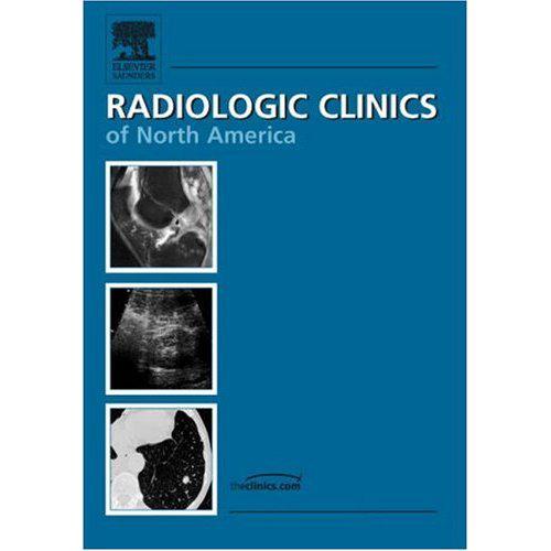
Radiologic Clinics Of North America Cardiac Imaging
Free Download
Volume: vol 42 Issues 3
Size: 10 MB (10931550 bytes)
Pages: 200/200
File format: pdf
Language: English
Publishing Year: 2004
Direct Download: Coming soon..
Download link:
Category: Medicine , Clinical MedicineSign in to view hidden content.
Be the first to review “Radiologic Clinics Of North America Cardiac Imaging” Cancel reply
You must be logged in to post a review.


Reviews
There are no reviews yet.